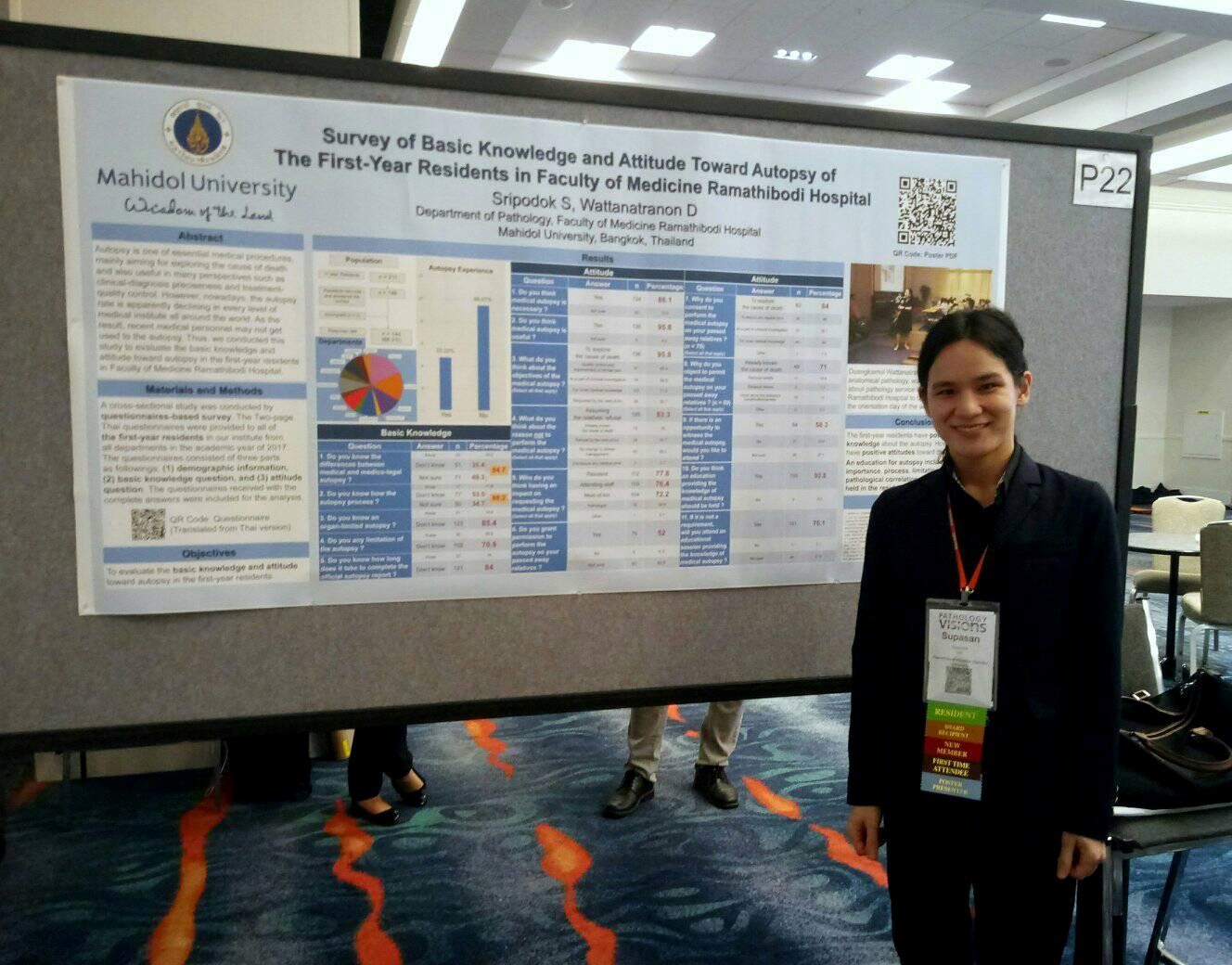
by Supasan Sripodok, MD, Anatomical Pathology Resident, Faculty of Medicine Ramathibodi Hospital, Mahidol University, Thailand; 2018 DPA Travel Award Recipient
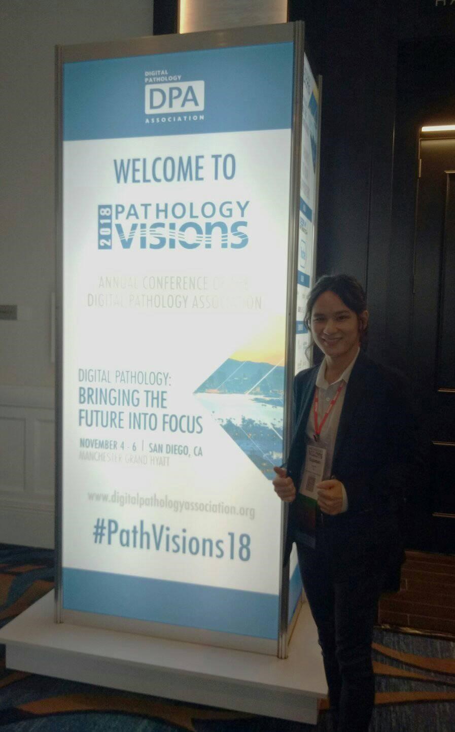
The 2018 Pathology Visions Conference was my first time attending a conference outside of Thailand. It was a huge international conference with more than 500 attendees from many countries and multidisciplinary specialists including pathologists, medical technicians, histologists, computer engineers and computer scientists, all participating in this collaborative conference. Many businesses associated with digital pathology exhibited their innovative and creative products including hardware, e.g. scanners, and software, e.g. computational artificial intelligence.
Before attending Pathology Visions 2018 in San Diego, whole slide imaging (WSI) was the only concrete aspect I knew about digital pathology. From my experience, the image files from WSI were used together with telecommunication/tele-pathology mainly for several inter-institutional conferences which were conducted three to four times per academic year. This showed that my knowledge regarding digital pathology was very limited.
Multiple educational sessions and workshops in the 2018 Pathology Visions Conference provided countless pieces of beneficial information opening my eyes to many more aspects of digital pathology. Many fascinating presentations given by the professional speakers were very informative and I had come to know many of the topics for the first time. The topic of artificial intelligence-assisted detection of nodal metastasis using deep-learning computation caught my attention. If this program is well trained and consistent, the tedious works of searching for nodal metastasis in a variety of cancerous tumors could be done faster than ever, helping to reduce workloads for pathologists.
Another session by Dr. Liron Pantanowitz, “Evolution of Digital Pathology = Revolution of Medicine” was stunning. He gave many examples of what pathologists and their work would be in the future if digital pathology becomes better developed and implemented in hospital workflow. For instance, we could do an intraoperative consultation for the frozen section anywhere, not just the hospital laboratory, by using tele-pathology. Finding the micro-organisms such as H. Pylori or acid fast bacteria would be more efficient with the help of artificial intelligence by matching them from a database. Deep learning-assisted programs would be able to help us in finding the abnormal cells in cervical cytology/Pap tests and help the cytopathologists to see the only noted cells instead of spending a whole day searching for pathologic cells. These mentioned examples will hopefully become a reality in the very near future.
Even though the technologies and innovations displayed in 2018 Pathology Visions conference seem to be promising, most of them are too advanced for the current utility in Thailand. In this developing country, we are still using the conventional work process, glass slides and microscopes to diagnose diseases. We have been using the traditional equipment for a long time, and nearly all of the pathologists have felt comfortable and confident using it.
In my opinion, the part of digital pathology that can be instantly applied for Thai conditions is integration into the curriculum for medical students as presented in the session of “Use Case for Digital Pathology with Tutor” by Dr. Douglas Hartman. For example, medical students learn the typical and characteristic features of some specific diseases as a representative sample of pathology. So, every academic year, the same sets of limited glass slides and the only need-to-know diseases are selected for teaching. However, the color of the slides fades as time goes by, requiring restaining or recutting of the slides to substitute the deteriorated ones. In order to solve this, using digital files instead of the glass slides is hopeful. Those files are permanent and don’t deteriorate. Another application that I can see the possibility, also for educational purposes, is to create collections of diseases for pathology residents. We could not only use them for reviewing before board examination, but also for many instances such as interdepartmental conferences, research purposes, and teaching sets.

Overall, Pathology Visions 2018 has totally changed my perception of digital pathology in a positive way. I am looking forward to future Pathology Visions Conferences, and I will surely encourage everybody who is interested and enthusiastic about digital pathology to attend. I guarantee that you will regard this as a special experience in your life.
Disclaimer: In seeking to foster discourse on a wide array of ideas, the Digital Pathology Association believes that it is important to share a range of prominent industry viewpoints. This article does not necessarily express the viewpoints of the DPA, however we view this as a valuable point with which to facilitate discussion.

Please log in to your DPA profile to submit comments