by Sibel Sensu, MD; Istinye University Medical Faculty, Pathology Department, Istanbul, Turkey
BACKGROUND
Telepathology, is sharing pathology data between distant computers using telecommunication technology (1). Medical education is considered a clerkship that is best accomplished by face-to-face interaction. However, in the changing world, hybrid education is inevitable as clearly seen in the COVID-19 crisis. For a long time, many alternative methods have been used for the execution of theoretical courses from distance (2-4). As well, there have been examples of online practical courses, reported from all over the world (5,6).
The microscopy practice course is a significant component of undergraduate pathology education which helps the student to visualize and comprehend deviations from normal to abnormal in cells and tissues (7-9). Although it has been performed mostly with traditional light microscopes; digital (virtual) microscopy is rapidly gaining importance (7,9,12-14). However, in the literature, there are very scarce reports on integration of digital microscopy to distance education (14,15).
In our university, microscopy practice courses of Pathology Department were realized in Digital Laboratory (DigiLab) since the beginning of the academic year, 2019-2020 (Figure 1). EduPath platform which is developed by Virasoft Inc. (Digital Pathology and Artificial Intelligence Software Inc.) was used for digital pathology microscopy education. EduPath software application is a web-based application that is based on WSI (Whole Slide Imaging) method. The users interactively login to the EduPath platform with the desired device (computer, tablet, smart phone etc.) at any location, whenever they prefer. In DigiLab, special usernames and passwords were assigned to users (students and educators), thus, they were able to access the scanned slides, analyze in low /high power as if they were looking through a microscope, and also annotate the images. In addition to high resolution images of the cases, clinical, radiological and laboratory findings of each patient were also uploaded to the EduPath. In this way, the system was helpful for case-based learning. ISO 27001 Information Security Management System and HIPAA compliance standards to which Virasoft was subject, ensured the data security. At the beginning of the academic year, 94 selected cases that were planned to be discussed during the academic year had been scanned with a special scanner and uploaded to the EduPath platform. As well, clinical and laboratory data along with gross macroscopic data had been added to each case. Personal passwords had been given to 2nd and 3rd-grade medical students.
In our university, distance education activities were performed via the support of the University Distance Education Center (UZEM) which used ALMS, the learning management system developed by Advancity. ALMS is a platform where students carry out all kinds of academic activities. On this platform, students can view the lectures assigned to them, access the materials added by lecturers in the course and communicate with relevant educators. Also, Perculus virtual classroom software owned by Advancity is integrated for real-time lectures on the ALMS platform. In Perculus virtual classroom software, the lecturer can interact with students by audio and video, share lecture documents, can make quizzes and use a whiteboard, as well as share the screen of her/his computer with students. UZEM had previously used to support for asynchronous execution of three obligatory courses in Medical Faculty (two language courses and a national history course). During the COVID-19 outbreak, distance education began in all theoretical courses of Medical Faculty on March 23, 2020, and the curriculum was completed on May 22, 2020, as pre-planned and announced. As well, the Digital Pathology Laboratory was performed remotely with the EduPath softtware application developed by ViraSoft Inc. We aimed to share and discuss our experience on the integration of digital microscopy to distance education since it is the first example in our country and one of the rare global examples.
MATERIALS AND METHODS
Participants
In the distance education period, distance education system of the University and the EduPath application were integrated to carry out the distance digital pathology microscopy course for 2nd and 3rd grade medical students (Figure 2).
Data Collection and Analysis
Three real-time pathology microscopy lectures were implemented (two with 3rd-grade and one with 2nd-grade medical students) between March 23, 2020, and May 22, 2020. Third-grade medical students were taking microscopy lectures from the beginning of the academic year and had already attended 8 hours of digital laboratory microscopy courses at the university; so they were used to and trained for the EduPath application. Pathology courses of 2nd-grade students began in April which coincided with the e-learning period due to the COVID-19 pandemic. For 2nd-grade students, a written guideline of the EduPath application was sent, and also, a trial course was done before the actual microscopy lecture. Then, the students accessed the cases shared with them by using their passwords at any time, any place and with the device (laptop, mobile phone, desktop etc.) they preferred to get ready for the real-time virtual microscopy course During the real-time microscopy courses, the lecturers and students came together using Perculus virtual classroom software on the ALMS-Perculus platform. Virasoft Edupath application was accessed by both lecturer and students and screen sharing was made by the lecturer to explain the cases. The students re-analyzed the cases with their educators and completed the courses interactively from distance, similar to the face-to-face courses at school.
After completion of the courses, feedbacks were obtained with an online survey (Google Form) which included the following questions: 1) How was the distance digital microscopy course? (5-point Likert scale; 1 very bad, 2 bad, 3 neutral, 4 good, 5 very good); 2) Did you take advantage of this practice? (5-point Likert scale; 1 none, 2 few, 3 neutral, 4 much, 5 very much); 3) What are the advantages of this system? (Open question); 4) What are the aspects of this system that need to be developed? (Open question); and 5) Should this system continue? (Yes/No)
RESULTS
One hundred sixty-eight medical students attended to real-time digital microscopy session for virtual Pathology class and 72 students (67.9%) approved and answered the online questionnaire. Respectively, 64% (n=14) of third grade students and 56% (n=27) of second grade students scored the practice as “very good”. For 14% (n=3) of third grade students and and 25% (n=12) of second grade students, the practice was “good”. The percentage of the students for whom the practice was “very bad”, was 0% for third grade students and 2.1% for second grade students (Table 1).
Respectively, 59% (n=13) and 23% (n=5) of third grade students found the system “very much” and “much” advantageous. For the second grade students, 46% (n=22) took “very much” advantage of the system while 33% (n=16), found it “much” advantageous. In other words, approximately 80% of the third and second grade students “very much and much” took advantage of this system. On the other hand, none of third grade and 2% (n=1) of second grade students did not take any advantage of the system (Table 2).
According to students, the advantages of the system were; “to analyze the cases whenever they want; to see the annotations of the educator; to annotate and ask questions; to repeat the analysis as much as they want and in detail”. Third-grade students stated that the distance microscopy practice was as good as the one done in school. According to students, the aspects needed to be developed were; “image quality and technical pitfalls”. It was thought that the image quality might be related to the internet connection speed of the institution and/or the student rather than the image itself. One third grade student noted that ‘though the lecture went on well, he preferred face-to-face practice’ (Figure 3). Finally, 83.3% of second grade and 86.4% of third grade students wished the system to continue.
The Cronbach Alpha reliability statistics score of the questionnaire used for the feedback survey was 0.966.
DISCUSSION
Digital pathology is a pathology practice performed by scanning the glass slide with a special high-resolution scanner; turning it into a digital slide and showing on a computer screen. It enables to annotate, archive and share the slide with others (1,16). Its value in routine medical practice is gradually increasing and also, a perfect digital pathology infrastructure is required for image processing and artificial intelligence applications in pathology (16). Digital pathology which is used for diagnosis, research, and consultation, has also a place in undergraduate and postgraduate education and training due to its many advantages over traditional light microscopy (7,9,12-14). First, since digital images are identical, each student has the opportunity to inspect the same good-quality image. Secondly, glass slides might fade, get broken, or lost in time while digital slides can confidently be stored. Thirdly, in tiny or rare tissues, to prepare one slide is enough for all students. Finally, digital slides can be analyzed repeatedly using computerized decision support systems and artificial intelligence applications; and, if necessary, a second opinion can be asked by teleconsultation. Last but not least, the EduPath platform is suitable for case-based learning as big data can be sent along with microscopic slides to enable the student to analyze the patient as a whole. Studies show that digital microscopy positively affects the learning process and communication skills of students. Moreover, it improves self-esteem of the students (7,9,12-14). However there are some pitfalls; It is always firmly emphasized that image quality should be high. Screen quality should also be optimal. Also, probable technical problems and low internet access rate have to be dealt with. Actually for a successful digital pathology practice, perfectly prepared, and perfectly-scanned slides, perfectly-working internet infrastructure, and high- quality screen are required (5). Digital pathology, when especially used for diagnosis, consultation, and research certainly require the highest quality. Undoubtedly, the same is required for upper-level teaching and learning activities.
Even though telepathology is rarely used for educational purposes, taking into account the current global conditions, it will undoubtedly gain significance. Telepathology used for education has several significant benefits. Especially during problematic conditions such as COVID-19 pandemic, for students in rural areas, for synchronous teaching to various remote centers, or for students with special needs or disabilities, online education might be crucial. Universities are facing increased budget constraints which lead to cost-effective solutions for laboratory activities. On the other hand coronavirus pandemic strikingly changed the education dynamics leading the invention and implementation of new learning / teaching models. In our university, based on the already established distance education platform, a very rapid transition to online education system was succeeded. In a very short time after the announcement of the first COVID-19 case (March, 11), at March 23, all faculties of our university began real-time online education. Distance histopathology courses were a very unique and successful solution for the continuence of Pathology curriculum which was to our knowledge, the first example in our country. This experience, after the pandemics, can be used as a part of hybrid education methods. It can be a solution for a better workforce allocation in medical schools with educator shortage, since in universities the academic members are also involved in heavy routine diagnostic activities. Also, in special conditions when the students are unable to attend school for a while, distance microscopy courses might be a supportive. Although there is a growing need for digital solutions, traditional microscopy should not be overlooked, as some students prefer face-to-face training and besides, hematology or clinical laboratory evaluations are still performed with traditional microscopy. But still, it is possible to establish an online histopathology lesson just similar to the one done in a face-to-face condition (17,18).
Sivamali (14), in a study done in Australia, Quennsland, James Cook University, School of Medicine and Dentistry, analysed the effect of online digital practical pathology approach on 4th and 5th year students. Fifty percent of the students found the practice important due to factors as high image quality, faster learning, convenience, technological advantages, high access rate and slide annotations. Internet availability in more remote locations and lack of quick adaptations to technology were mentioned as the problems. In a study by Zhong from China (19), during COVID 19 pandemic, 192 third year dental students had an online virtual microscopy experience. As a result, application of remote learning and virtual micrsocopy were effective on histopathology learning. Another current study has been published from Israel by Samueli et al. (20). In that survey, 25 students who answered the anoymous questionnaire found the remote digital histopathology course very useful. They mentioned that, they understood the covered diseases better, and they strongly recommended the course to others. They added that the most significant disadvantage was technical challenges. Similar to previously published studies (14, 19, 20), our survey showed that a high percentage of students were satisfied with the online digital microscopy course experience. However, our analysis also showed that the technical problems, internet availability, and quality of the virtual slides were critical issues to be handled during digital pathology and telepathology, as mentioned by others (9-14).
This survey was based only on students’ perceptions and opinions on online digital microscopy experience, which, according to us, was one of the limitations. Thus, a subsequent survey is planned to combine these results with an assessment on pre- and post-course performances of the participants. Another limitation was the difference of experience between the second and third grade students on telepathology. We found that third grade students were more satisfied with the courses than the second grade students. Since second grade students had met the system for the first time while third grade students had a previous face-to-face experience in school, this difference of experience might have led to the distinct satisfaction rates. Therefore, the subsequent survey might give additional and reliable data.
CONCLUSION
Medical education is a discipline where it is best if the educator and learner are face-to-face. However, in the developing and changing world, distance education provides support and convenience. The integration of e-learning to both theoretical and practical areas of medical education is a subject to be developed by a team composed of health, computer and education experts. COVID-19 pandemic inevitably led to many solutions in the establishment of various aspects of medical education. The digital pathology microscopy infrastructure of our university and integrating it into the e-learning system helped us carry on the microscopy lectures similar to the ones performed at school. This is the first and for now the only experience in our country, and one of the few global examples where remote digital microscopy application is used in digital pathology education. We believe that this experience will contribute to raising doctors ready for the digital and artificial intelligence-based applications of the future.
Figure 1. Pathology microscopy course in ISU Medical School DigiLab
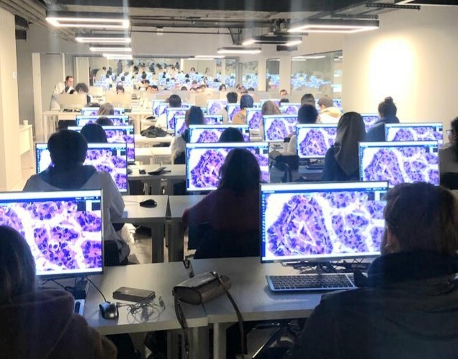
Figure 2. Integration scheme of the Virasoft’s EduPath platform and distance education system
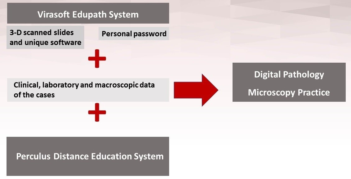
Figure 3. Advantages of the digital microscopy system in the distance digital pathology course and the parts that need to be improved according to students
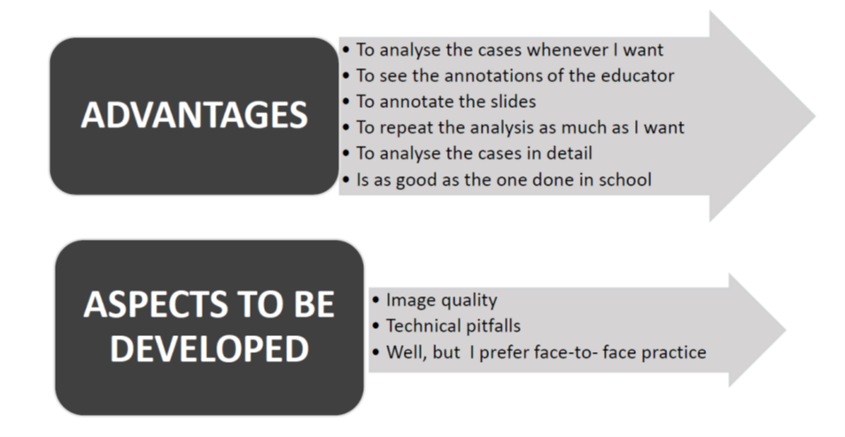
Table 1. Opinions of second and third grade students on the remote digital microscopy experience during virtual Pathology class ( How was the distance digital microscopy course?)
|
|
Third grade N(%) (n=22) |
Second grade N (%) (n=48) |
|
Very good |
14 (63,6) |
27 (56,3) |
|
Good |
3 (13,6) |
12 (25) |
|
Neutral |
4 (18,2) |
6 (12,5) |
|
Bad |
1 (4,5) |
2 (4,2) |
|
Very bad |
0 |
1 (2,1) |
Table 2. Opinions of second and third grade students on the remote digital microscopy experience during virtual Pathology class (Did you take advantage of this practice?)
|
|
Third grade N(%) (n=22) |
Second grade N(%) (n=48) |
|
Very much |
13 (59,1) |
22 (45,8) |
|
Much |
5 (22,7) |
16 (33,3) |
|
Neutral |
3 (13,6) |
6(12,5) |
|
Few |
1 (4,5) |
3(6,3) |
|
None |
0 |
1(2,1) |
Authors:
Sibel Sensu1, Ozgur Teke2, Muammer Demirci3, Sevcan Kutlu1, B.Nazlı Genc1, Samet Ayalti2, Yesim Saliha Gurbuz1, Nusret Erdogan1
Affiliations:
(1)Istinye University Medical Faculty, Pathology Department, Istanbul, Turkey
(2) Virasoft Digital Pathology and Artificial Intelligence Software Inc., Istanbul, Turkey
(3) Istinye University Distance Education Center, Istanbul, Turkey
REFERENCES
1-Barisoni, L., Gimpel, C., Kain, R., Laurinavicius, A., Bueno, G., Zeng, C et al. Digital pathology imaging as a novel platform for standardization and globalization of quantitative nephropathology. Clin Kidney J 2017;10(2): 176–187.
2-Newman, NA., Lattouf, OM. Coalition for medical education-A call to action: A proposition to adapt clinical medical education to meet the needs of students and other healthcare learners during COVID-19. J Card Surg 2020; 35(6):1174-1175.
3-Pei, L., Wu, H. Does online learning work better than offline learning in undergraduate medical education? A systematic review and meta-analysis. Med Educ Online 2019; Dec;24(1):1666538.
4-Sandhu, P., de Wolf, M. The impact of COVID-19 on the undergraduate medical curriculum. Medical Education Online 2020; 25, 1764740. https://doi.org/10.1080/10872981.2020.1764740
5-Brockman, R.M., Taylor, J.M., Segars, L.W., Selke, V.& Taylor, T.A.H. Student perceptions of online and in-person microbiology laboratory experiences in undergraduate medical education. Med Educ Online 2020; Dec;25(1):1710324.
6- Donkin R, Askew E, Stevenson H. Video feedback and e-Learning enhances laboratory skills and engagement in medical laboratory science students. BMC Med Educ 2019; Aug 14;19(1):310.
7-Lee, B.C., Hsieh, S.T., Chang, Y.L., Tseng, F.Y., Lin, Y.J., Chen, Y-L et al. A Web-Based Virtual Microscopy Platform for Improving Academic Performance in Histology and Pathology Laboratory Courses: A Pilot Study. Anat Sci Educ 2019; 10.1002/ase.1940. Advance online publication. https://doi.org/10.1002/ase.1940.
8-Sagol, O., Yorukoglu, K., Lebe, B., Durak, M.G., Ulukuş, C., Tuna, B, et al. Transition to Virtual Microscopy in Medical Undergraduate Pathology Education: First Experience of Turkey in Dokuz Eylül University Hospital. Turk Patoloji Derg 2015; 31(3):175180.
9-Simok, A.A., Hadie@Haji, S.N.H., Abdul Manan@Sulong, H., Yusoff, M.S.B., Mohd Noor, N.F., Asari M.A et al. The impact of virtual microscopy on medical students’ intrinsic motivation. Education in Medicine Journal 2019; 11(4):47–59.
10-David, L., Martins, I., Ismail, M.R., Fernandes, F., Sidal, M., Seixas, M et al. Interactive Digital Microscopy at the Center for a Cross‑Continent Undergraduate Pathology Course inMozambique. Pathol Inform 2018; 9:42.
11-Foad A.F.A. Comparing the use of virtual and conventional light microscopy in practical sessions: Virtual reality in Tabuk University. Journal of Taibah University Medical Sciences 2017; 12(2), 183e186
12-Nauhria, S., Ramdass, P. Randomized cross-over study and a qualitative analysis comparing virtual microscopy and light microscopy for learning undergraduate histopathology. Indian J Pathol Microbiol 2019; Jan-Mar;62(1):84-90.
13-Samal, N., Prakash, R.V. Randomized cross-over study and a qualitative analysis comparing virtual microscopy and light microscopy for learning undergraduate histopathology. Indian J Pathol Microbiol 2019; 62:84-90.
14-Sivamalai S, Murthy S.V, Gupta T.S, Woolley T. Teaching pathology via online digital microscopy: positive learning outcomes for rurally based medical students. Aust J Rural Health 2011; 19(1):45-51.
15-Weinstein R.S, Graham A.R, Richter L.C, Barker G.P, Krupinski E.A., Lopez A.M et al. Overview of telepathology, virtual microscopy, and whole slide imaging: prospects for the future. Hum Pathol 2009; 40(8):1057-1069.
16-Niazi M.K.K, Parwani A.V, Gürcan M.N. Digital pathology and artificial intelligence. Lancet Oncol 2019; 20: e253–61.
17- Hamilton P.W., Wang Y, McCullough S.J. Virtual microscopy and digital pathology in training and education, APMIS 2012; 120, 305-315
18- Saco A, Bombi JA, Garcia A, Ramírez J & Ordi J. Current status of whole-slide imaging in education. Pathobiology 2016; 83, 79–88.
19- Zhong Y, Sun W, Zhang W, Liu L, Xu Y & Jiang Y . Application of Remote Online Learning in Oral Histopathology Teaching: An Acute Response to the COVID-19 Pandemic. (2020, Preprint). DOI: https://doi.org/10.21203/rs.3.rs-51823/v1
20- Samueli, B., Sror, N., Jotkowitz, A., & Taragin, B. Remote pathology education during the COVID-19 era: Crisis converted to opportunity. Annals of Diagnostic Pathology 2020; 49, 151612. Advance online publication. https://doi.org/10.1016/j.anndiagpath.2020.151612 .
Disclaimer: In seeking to foster discourse on a wide array of ideas, the Digital Pathology Association believes that it is important to share a range of prominent industry viewpoints. This article does not necessarily express the viewpoints of the DPA, however we view this as a valuable point with which to facilitate discussion.
2 comment(s) on "Telepathology in Medical Education"
02/11/2021 at 03:50 PM
Diane Kenwright says:
Great article. It would be really helpful if we could see the virtual images. Is it possible to access them?04/17/2021 at 05:29 AM
Sibel Sensu says:
Dear Diane Kenwright,Thanks for your comment. For now, the digital slides can only be accessed with a private password given by the company, Virasoft. Also, the slides belong to the university which also is a second issue that they are unfortunately not widely accessable.
If I can have a direct contact with you, I would like to find a solution to introduce the system to you.
Here is my e-mail:
sibel.sensu@istinye.edu.tr
Sincerely,
S.Şensu
Please log in to your DPA profile to submit comments