by Jonhan Ho, MD, Assistant Professor, UPMC
Many months ago, through luck and maybe a little destiny, a 14-year-old, no longer used high throughput slide scanner came to my attention. I petitioned to see if we could have it for research and educational purposes. After some heavy lifting (literally and figuratively), we were gleefully making arrangements to have it picked up and delivered. We decided to drop it right in the middle of our sign out room.
Our sign out room is communal, like the living space of a college dorm suite. We have two multiheaded microscopes that the attendings use for their daily signout, and 4 individual microscopes for our two dermpath fellows and any rotating dermatology or pathology residents to use. At any one time there are typically 4-6 attendings and trainees “slapping glass” on the microscopes in the room. Additionally, our lab staff pick up, drop off, and match up hundreds of slides per day to our mailboxes. We get lots of work done in that room every day, but most importantly, we exchange ideas and talk about interesting cases. Our sign out room is the central hub of activity in our dermatopathology division.
We had a choice of two locations to put the scanner - in the histology lab which was just down the hall from the sign out room or in the sign out room itself. Since the scanner couldn’t be hooked up to the network without significant effort, we decided to put the scanner right smack dab in the middle of our sign out room instead of having someone trudge back and forth with USB sticks. I wasn’t sure how often it would be used, so I briefly and rudimentarily trained my fellows and colleagues on how to operate the scanner in a de-identified fashion.
The fellows were the most excited about having the scanner. Each fellow is responsible for one unknowns conference per week, 10 cases per conference. Before we acquired our scanner, the fellows used a scanning service outside our division, which required the cases to be packed up and sent a week before the conference. After we acquired the scanner, the turnaround time was nearly completely eliminated. However, once they got a taste of having the scanner at their disposal, there was no going back. The fellows started scanning all the interesting cases they came across for their own collections. When a rare case came across the signout service, they would pop it right in the scanner. Since we were all in the same room, a fellow walking over to the scanner would catch interest from others in the room. “Whatcha got there?” a rotating resident would ask. “Got something interesting?” another attending would ask. The simple act of walking over to the scanner with a case in hand became the trigger for discussion about the case and the disease and then before you knew it, ideas for research projects or papers would be born.
As more and more scanning was performed, the allure of whole slide images began to diffuse outside of our division. The dermatology residents began to ask to see interesting cases. Dermatology faculty began to ask for scans to be presented at conferences. Our Mohs surgeons in particular were very interested in seeing the whole slide images at the weekly Mohs clinicopathological conference so we created a workflow in which they would request cases and the fellows would scan them. Image requests from the dermatologists and from other departments became much easier to handle. In a unique syphilis case, our fellow scanned a case and presented the images at the infectious disease conference, and the medicine department was so impressed with him that they invited him as a guest lecturer on dermatological manifestations in HIV patients. While the fellow’s knowledge was the reason for the invite, the ease of acquiring images allowed the fellow to respond quickly and with high quality. It wasn’t just the fellows that started using it - our faculty wanted their own whole slide images too. Two faculty would scan their own cases, and a third would ask the fellows to scan it for them. A poll of the faculty showed that they scanned for a few key reasons: (1) interesting cases, (2) research/presentations/publications, (3) conferences, and (4) teaching. We all used a cloud-based platform to help organize, access, and share our whole slide image collections.
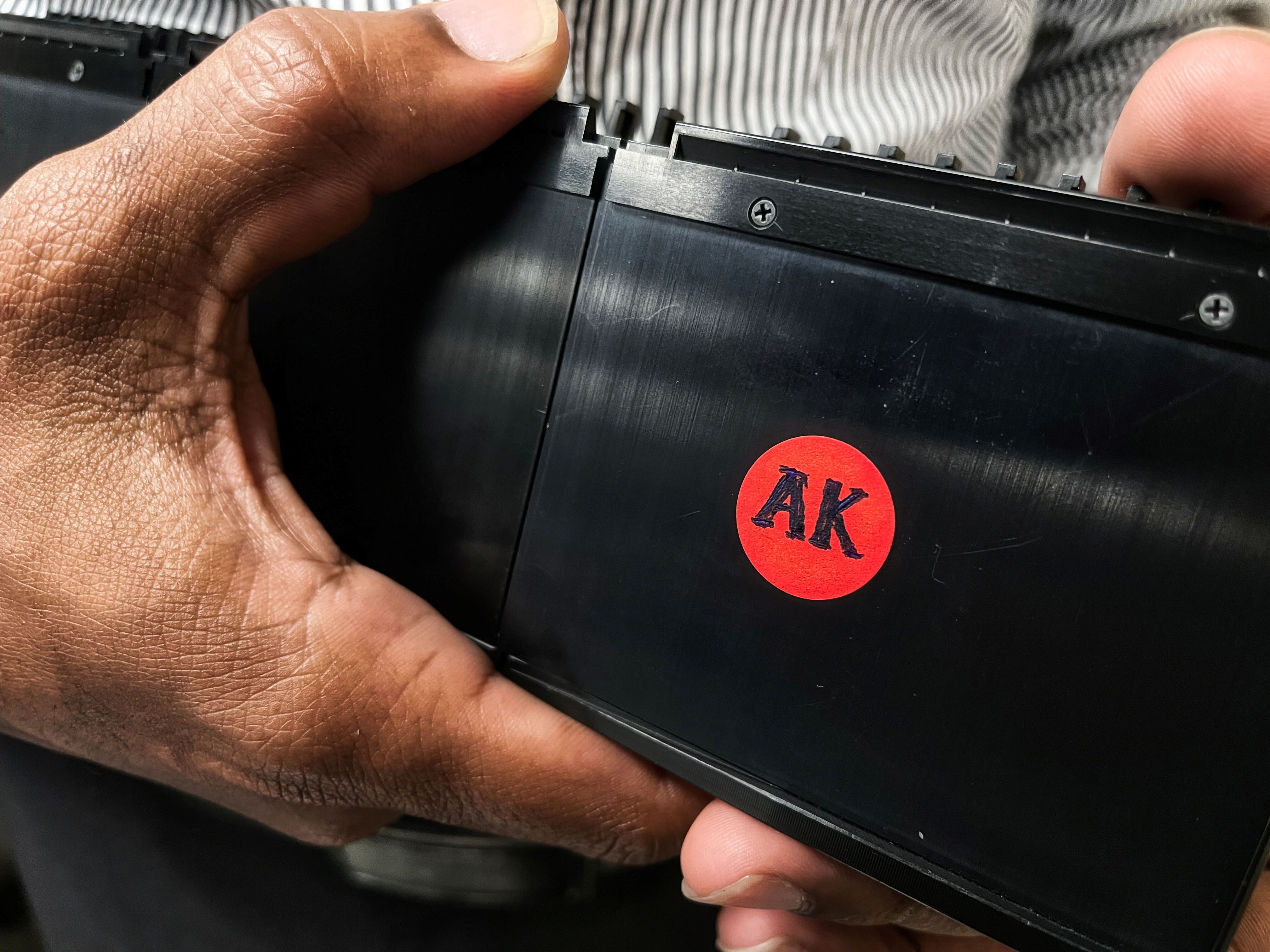
There were some issues that we had to work through. The scanner became so popular that people would come in looking to scan their own slides only to find that the scanner was occupied. Since the scanner is a batch scanner, new batches could not be loaded and put in the queue without disrupting the current batch and the user would have to keep checking back for availability. Some newer scanners now have continuous loading designs with hot swappable magazines to alleviate this issue. Since it takes some time to scan the slides, if the batch completed and another user wanted to scan the slides, they would remove the magazine and place it on top of the scanner. This quickly became an organizational issue, and we have since implemented a one magazine-one user workflow. Each user gets their own magazine, labeled with their initials, so we know where the slides should go after scanning. A few months ago, the light source fan failed (after 14 years!) and the scanner was down for a couple weeks while waiting for the part to arrive from Japan.

The settings that we found worked best for research and education were those that prioritized image quality over scanning time. We set the scanner to place more focus points and used smaller tiles, which significantly increased the time it took to scan each slide. To alleviate this, sometimes we would scan manually, picking only one of the sections on the slide to scan instead of all of them. Since we typically have four sections per slide, this cut down the scan time and file size by 75%, but at the cost of needing to monitor the machine. I wish that there was an “education mode” - an artificial intelligence algorithm to identify and scan just the best section instead of all of them. And while we knowingly prioritized image quality over scan time, we acknowledge that since there was often a queue for the scanner, faster focusing algorithms and scanning would help our specific workflow. Immunohistochemical stains in particular were difficult to get focused uniformly. Tissue on IHC slides is typically thicker than on H&E slides and thus more difficult to cut on the microtome, especially so on small sections such as seen in skin specimens. These sections are more prone to folds and chatter, and coupled with a weaker counter stain, give default focus algorithms some trouble, an issue that affects all scanners to some degree. It is likely that we could at least partially rectify this issue by creating a custom scanning template with different sensitivities specifically for IHC slides, but we just haven’t been able to dedicate the time to do this. Furthermore, we often mix and match H&E and IHC slides within a batch to keep a case together, and in batch mode there is no way to indicate per slide settings that we know of. So, for my second wish, I’d like the scanner to use AI to know what type of slide it is and apply the best settings accordingly.
Today the scanner feels like one of us. We feel its presence every day. We see it when we come in, we interact with it several times per day, and while we are cranking out cases, it is also hard at work cranking out whole slide images. Dozens of slides pass over its stage every day. Even when we are not directly interacting with it, its constant whirring of activity and endearing robotic noises remind us that it is busy helping us as best it can. The sounds it makes tell us if it is loading a slide, running through focus points, actively scanning a slide, or returning the slide to the magazine. It reminds me of a Roomba (albeit much more sophisticated!), a happy, reliable robot that serves us every day with essential and important tasks to the best of its ability, never even asking for anything as much as a thank you.
Perhaps most telling is what my fellow said when they graduated at the end of their fellowship. I asked him what he will miss most. He said, “It’s going to suck not being able to scan my cool cases.” It’s hard for me to imagine life before our friendly neighborhood slide scanner.
Disclaimer: In seeking to foster discourse on a wide array of ideas, the Digital Pathology Association believes that it is important to share a range of prominent industry viewpoints. This article does not necessarily express the viewpoints of the DPA, however we view this as a valuable point with which to facilitate discussion.
5 comment(s) on "We put a slide scanner right in our main sign-out room. Here’s what happened."
07/09/2021 at 12:38 AM
Colleen Beatty says:
Great read.07/09/2021 at 01:10 PM
Winston Low says:
We supplied Whole Slide Images (WSI), digital imaging scanner, 20 slides per magazine with image resolution save at 1X, 5X, 1, 2, and 4. Can be annotated remotely.07/11/2021 at 03:40 PM
Jonathan He says:
Go Digital!07/12/2021 at 10:03 AM
Daniel Gonzalez says:
Excellent read. As scanners become more accessible to the everyday pathologist, I see them being used more frequently. It's analogous to the adoption of computers over the years. They were once these large, expensive devices that were only used for bleeding edge technologic applications and now it's hard to imagine life without them.10/30/2021 at 12:49 PM
Caroline Miller says:
Interesting read, thank you.Please log in to your DPA profile to submit comments